Nu-Venture LLC
TRICHOMES FROM THE KINSMAN PROJECT OF 18" X 24" FLATS
TRICHOMES FROM THE KINSMAN PROJECT OF 18" X 24" FLATS
Couldn't load pickup availability
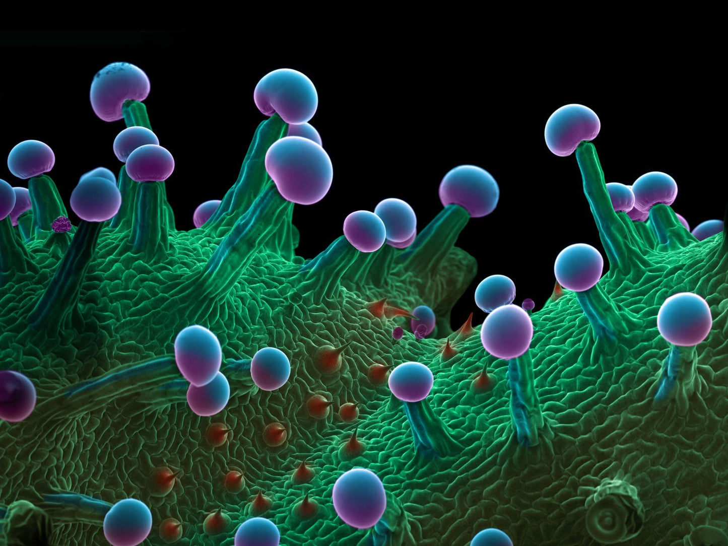
Art meets science in this uniquely stunning photograph that reveals the extraordinary microscopic beauty of the world’s most controversial plant, cannabis sativa.
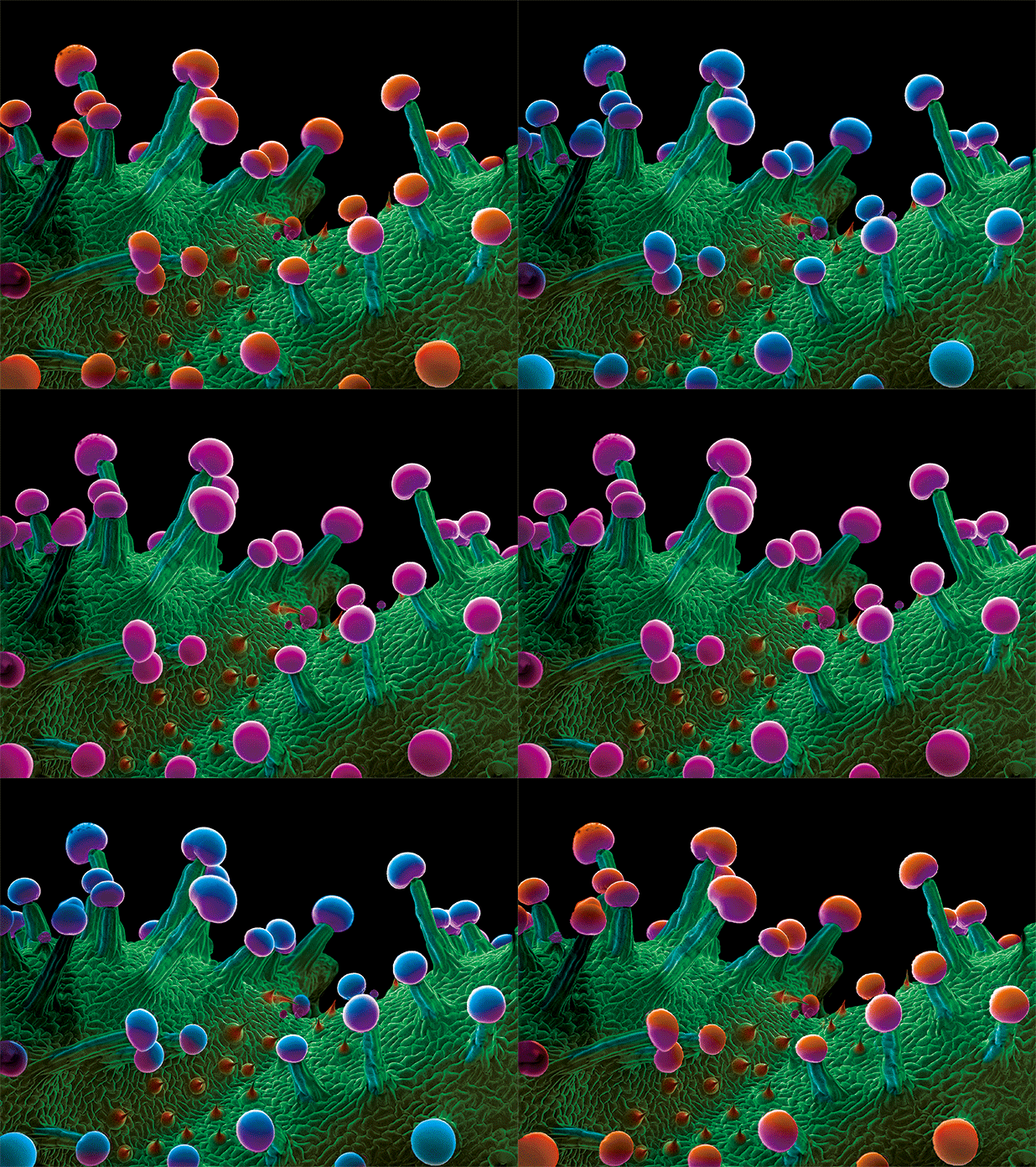
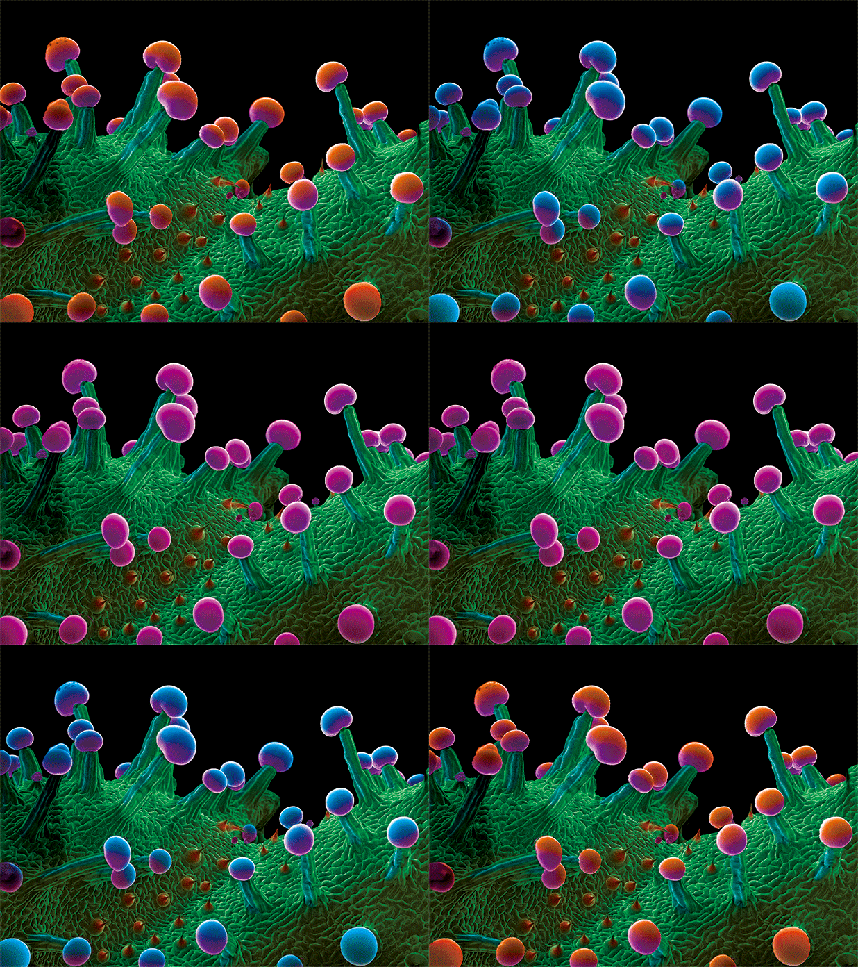
This color-enhanced scanning electron micrograph (SEM) image of the surface of a marijuana plant leaf shows glandular cells, called trichomes. These are capitate trichomes that have stalks. They secrete a resin containing tetrahydrocannabinol (THC), the active component of cannabis. The spherical cells at the top of the trichomes are 60 micrometers in diameter. Our artwork is a 3-flip design with the colors of the caps changing as you change your perspective.
The Kinsman Project
Author of "Marijuana Under the Microscope", Ted Kinsman is an associate professor in the Photographic Sciences Department at Rochester Institute of Technology in Rochester, New York. He teaches high-speed photography and scanning electron microscopy, (SEM), while also holding degrees in optics, physics, and science education.
Super High-Resolution Print Creates a Crisp Image.
Hang this artwork in your home, office, dispensary or grow room for a great conversation starter.
The picture is a wrapped flat suitable for framing.
Measures 18" x 24". Weighs about 1 lbs.
NOTE: It is difficult to show a 3D product in a 2D format.


What customers are saying.....


